Lumbar Compression Fracture, Illustration - Stock Image - C027
4.6 (150) · $ 14.99 · In stock

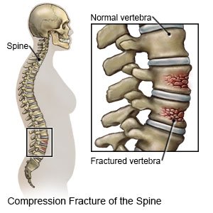
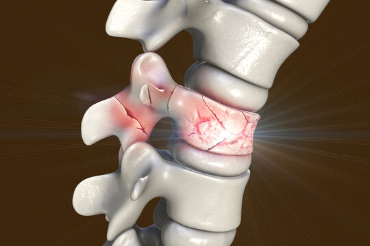
An interpretive illustration of an MRI depicting a sagittal view of compression fractures at the L1 and L2 vertebrae as a result of osteoporosis. Over time as bone becomes weaker and more porous, they become more susceptible to injury and fractures, especially in situations where significant weight or stress is placed on the bone. Evan Oto/SCIENCE PHOTO LIBRARY
Virginia Interventional Pain & Spine Centers
127 Vertebral Compression Fracture Stock Photos - Free & Royalty

Thoracic vertebral collapse, X-ray - Stock Image - C038/6661
110+ Compression Fracture Spine Stock Illustrations, Royalty-Free

Compression Fracture Stock Illustrations – 213 Compression

110+ Compression Fracture Spine Stock Illustrations, Royalty-Free
Compression Fracture Images – Browse 2,195 Stock Photos, Vectors

Lumbar Compression Fracture, Illustration - Stock Image - C027/6314 - Science Photo Library

Spinal Compression Fractures Advanced Orthopaedics & Sports
Compression fracture with signs of PLC injury in two patients. (a